


Among the diseases of the musculoskeletal system, osteoarthritis is a frequency leader.It is believed that the vast majority of the population of the planet at the age of 60 has the initial signs of changes in the joint cartilage and 14% have already manifestations of osteoarthritis.The most common version of this disease is the osteoarthritis of the knee joints.
However, "arthrosis" or "arthritis"?
Don't mix these two concepts.Arthosis is the process of change mainly of the structure of the joints and arthritis is an inflammation that could occur both against the background of the "intact" structure and on the background of arthrosis.
The changes in the bones with arthrosis can be compared, for example, with subsidiaries knotted on a tree trunk, which grows near the concrete fence and puts pressure on this fence with all its weight.
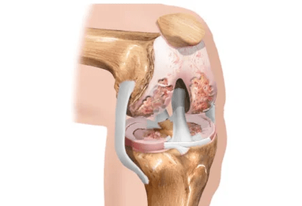
Normally, the surface of the bones once the other is separated from two layers of cartilage and Meniscis (additional cartilaginous plates).In addition to the role of the "buffer" between the bones, the cartilage provides the bone platelet and mechanical correspondence between them.The meniscus, which, due to large or small (but frequent) lesions and also lose their elasticity, can break completely or partially even more.
With age, and especially in the presence of a hereditary predisposition, the joint cartilage is thinner.This is the reason why the bones of the hips and the lower legs, which make up the articulation of the knee with their ends, are dangerously approaching each other, friction can even arise with each other.
Usually in parallel with the thinning of the cartilage over the years, another unpleasant event occurs: the amount of intra -articular fluid decreases.This liquid is not only a purely mechanical "lubrication" of the joint from the inside.It provides bone nutrition, menisci and joint cartilage.The violation of the "supply" of all these structures is a real disaster for the joint!
If there is a physical overload of the joint, bone extensions appear on the surfaces of the bones and begin to grow, more similar to the pointing or peaks.For the knee joint, these overloads will be the lifting of weights (including the overweight of your own body!), Physical work with an emphasis on the knees (for example, deserting the garden), constant walking on the stairs, running, wearing uncomfortable shoes, flat feet and many others.Now it is easy to imagine what is happening inside the knee joint during the development of arthrosis and how it manifests itself in appearance.
How does the relative work?
Each of us has seen the joint cartilage many times at the end, for example, chicken bone.It covers small areas of contact with bones.Under the joint cartilage there is a subcondic or pericilated bone.The human musculoskeletal system is arranged similarly.
Most of the person's joints are made up of bones, synovial shell (joint) and intra -articular fluid.
What happens to the articulation with arthrosis?
Under the influence of all those loads that have already been mentioned, there is a compaction and growth of the thin bone, following this, has increased the trauma of the joint cartilage.
The products of the cartilage cartridge formed due to the microtrauma fall within the synovial fluid.It is so organized by nature that they are foreign substances for the synovial shell and cause its inflammation.The formation of the synovial fluid is disturbed, which is usually a sort of "conveyor", similar to a continuous cycle of enrichment and purification of the blood.In addition, the joint fluid becomes lower than hyaluronic acid.It is worth saying this acid.
Hyaluronic acid provides the viscosity of the synovial fluid, creates the "buffer effect" and the "lubrication effect" between the bones, reducing their friction against each other.It is thanks to this substance that the articular fluid in the consistency recalls the egg protein, not water.Another important role of hyaluronic acid is to guarantee the delivery of nutrients from the articular fluid in the joint cartilage, since there is no place to bring nutrition: the blood vessels are not directly suitable for cartilage.Likewise, the "expenses" substances from cartilage to joint fluid are removed: using hyaluronic acid molecules.
Therefore, an improved bone seal occurs and unbearable conditions for joint cartilage are created.
The cartilage receives a signal to adapt to these extreme conditions and its change begins, in another way in which it is called remodeling.This manifests itself mainly from a decrease in cartilage elasticity.
In the advanced phase of the development of arthrosis, the bone becomes rigid, but at the same time more fragile, the cartilage itself is partially impregnated with calcium - calcified.
Symptoms
The development of osteoarthritis begins with a slight knee pain, which appears after walking through the stairs, physical activity, long walking on foot.Such a mild pain may appear for several months, or even years.So they become more pronounced.In the initial phase of the development of the disease, the knee bones are not deformed, but a light swelling of the articulation itself can be observed.
In the second phase of the development of the disease, the pain becomes more intense and occurs after a slight load.In addition to the pain, a crunch appears in the knee joint, which differs from the usual soft abuse of a healthy articulation with pain.Furthermore, the deformation of the joint becomes evident, the bones to the touch become wider and rude.Custe the knee more than 90 degrees becomes problematic.
In the third phase of the disease, knee pain becomes serious and constant, not even going through the rest period.The mobility of the knee becomes minimal, often it does not fold more than 90 degrees and does not extend to the end.The deformation of the bones of the joint becomes so strong that there is a curvature of Valgus (in the shape of an X) or varability (in the shape of O) of the legs.
Diagnostics
Inspection
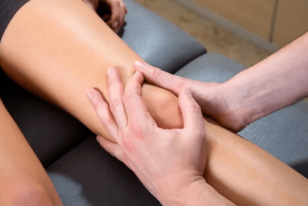
In the early stages of the disease, the joint has not changed, mobile, the muscles around it are preserved and strong enough.Only from the palpation (pressure) of some points, more often on the internal surface of the joint, the local (local) pain is determined.The doctor asks the patient to perform several squats, bending, straightening the leg on his knees, puts his face on the sofa and leads by himself to the flexion extensions (this is called "passive" movements).In this case, in addition to the pain and limitation of the volume of movements, it is possible to determine the creaking, by clicking on the joints.With a pronounced inflammatory component, the articulation is increased size, it seems that it is "pumped" with liquid.With a distant diffusion process, the flexion of the knee can be partially or completely absent, when examined, the surface of the articulation seems irregular, tuberous, the limb can be curved (movement of the axis of the limb, "evoked").
Laboratory and instrumental search
- The mandatory laboratory survey program includesGeneral blood tests, biochemical and immunological, urine analysis.In the examination of the general blood, attention will be paid: the increase in the level of leukocytes and the increase in the rate of settlement of erythrocytes, which indicates inflammation.In the biochemical analysis of the blood, metabolic metabolic indicators are important, the level of "liver enzymes".In immunological analysis, the presence or absence of signs of systemic inflammation will be determined, this is highlighted by the level of c-reactive protein.Urine analysis will reveal the content of "sand" - uric acid crystals.
- Analysis of the synovial fluid (joint)It is prescribed if this liquid is in sufficient quantities.That is, when the joint is swollen, swollen.In conditions of conformity to sterility, the doctor pierces the joint capsule in a strictly defined place, inserts the needle into the joint cavity and therefore removes the excess fluid.Part of the material obtained enters the laboratory for the analysis.At the end of the procedure, the anti -inflammatory drug of the glucocorticosteroid group is often administered in the joint cavity (for example, Diprospan).
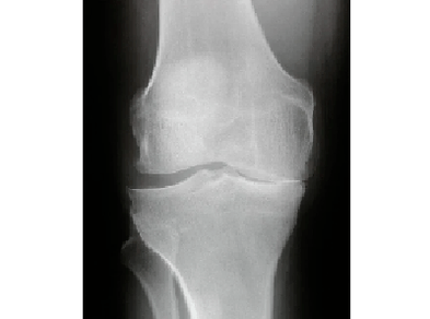 radiography.An image of both knee joints is mandatory, this is necessary to compare a sick knee with a healthy one.In the context, the attention is paid to the width of the joint gap (it is judged by the state of the meniscus and cartilage), by the presence or absence of bone-osteophytes peaks, signs of destruction (destruction) of the bones.
- Ultrasound of the knee jointsHe will answer questions about the conservation of the meniscus, on the presence of a baker cyst, the severity of inflammation, the presence or absence of uric acid crystals (in the presence of gout).
- MRI (magnetic resonance imaging).This study is prescribed if an ultrasound does not give an exhaustive answer to the questions of a specialist.Magnetic resonance imaging is mandatory for those patients who plan to conduct arthroscopy.
- Arthroscopy.It allows you to view, that is, personally evaluate the conditions of the joint.The method is essential for controversial diagnosis, suspicion of traumatic damage to Meniscis and ligaments (therefore directly during the study, it is possible to quickly remove the meniscus or torn ligaments).
Treatment of arthrosis of the knee joint
The principles of complete treatment should be followed, which include:
- Detailed awareness of the patient on the disease
- The use of physiotherapy exercises, which includes: specific exercises for the joints in the lying position, swimming
- Maintain an optimal body weight
- Wear a swing (soft bandage or at least an elastic bandage) during an increase in the load on the joint - on the road, during a walk and so on.
- Non -kerarative methods (physiotherapy).This type of treatment provides excellent results with the arthrosis of the knee joint (gonartrosis).Apparently, this is due to the fact that the articulation is available for the influence of factors such as magnetic and laser radiation.To treat the knee joint, it is possible to use magnetic currents, UHF, Crio exposure (translated from the Greek means the effect of the cold).The physiotherapy procedures are widespread, the treatment courses are generally short -lived - 10, maximum sessions per day or every two days.It should only be remembered on possible contraindications, including tumor processes, thyroid gland diseases and pelvic organs, as well as systemic inflammatory diseases (autoimmune).
- Drug therapy.

Principles of therapy with osteoarthritis:
- Relieve pain
- delay the further destruction of joint structures
- Restore the function of the lost articulation.
Non steroidal anti -inflammatory drugs
For pain relief, fans fans drugs are used -non -poundic anti -inflammatory drugs are used.Are used in and in the form of applications (application to the skin).Applications (local therapy) are a very effective method, especially when it comes to the early stages of the disease.Before using a gel or a cream containing fans, it is necessary to make sure that there are no changes on the skin, whether they are rash, pustules or cracks.The general rule of local treatment is to use the cream or the gel selected at least twice a day and if unpleasant sensations arise - to cancel the complete disappearance of these events.The intramuscular administration of painkillers is not currently recommended, since the risk of side effects following the administration using a syringe does not decrease, but rather the opposite.In the case of the pronounced inflammation, the accumulation of a large amount of glucocorticosteroid drugs is allowed, but it should be noted that this procedure should not be performed no more than 1 time every 3 months.
Condroprotectors
An anti -inflammatory effect higher with osteoarthritis are chondroitin or glucosamine preparations.They, like fans, fight with inflammation at the level of thin joint structures, but have fewer side effects and, above all, maintain their anti -inflammatory effect several months after cancellation.
Condroprotectors are a collective name for a group of drugs containing the condroitin sulphate and glucosamine - "construction bricks" of the cartilage.Despite the apparent high cost treatment with chondroprotectors, their convenience for patients and efficacy is difficult to overcome.Firstly, these substances, accepted inside, are perfectly absorbed by the stomach and the losses of the drug "along the road" to the cartilage are minimal.Secondly, they are able to suppress inflammation in the joint and, moreover, reliably slow down the process of destruction of the joint cartilage!Very often they have prescribed courses, because they have a "rather long ace that lasts several months and sometimes even up to six months.
The drugs based on hyaluronic acid are the ialuati so called.These funds are sold in the form of syringes prepared for intra -articular administration.Hyaluronic are an artificial synovial fluid.The effect of treatment with this method can last up to 12 months.
Surgical treatment of arthrosis of the knee joint
As for the arthrosis of the hip joints, in the case of serious changes and persistent function loss, it reaches the operation.With Gonartrosis, two types of interventions are currently performed: arthrodesis (motionless compound) and endoprothetics.The first operation is rarely performed, according to special indications, when the installation of an endoprotesis is impossible for any reason.The result of this operation is that the knee becomes immobile.But it doesn't hurt.The endoprothetic operation is much more profitable in terms of function.Recall that, with a great body weight, this operation is not performed: the risk of complications in the postoperative period is too large.From the moment of removal of the damaged sections of the articulation and installation of the prosthesis until the function is completely restored, more than three weeks pass.
How can it threaten ridiculous osteoarthritis?
Over time, osteoarthritis does not go back, but only worsens, above all while maintaining provocative factors.Consider the main sources of danger to the health and life of a patient with osteoarthritis.
- Chronic pain of various intensity- A very important risk factor, especially in the elderly.Constantly experienced unpleasant sensations can lead to sleep disorders, a reduced background of mood and even depression.It is difficult to predict which a adverse chain of events attracts the listed phenomena.
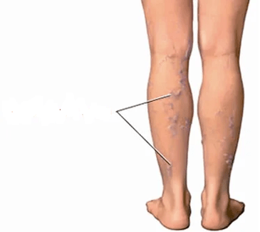
- Pathology of the veins.Constant inflammation in the knee area, the growth of bone-osfitite peaks, which can mechanically hurt the poplitei vessels, can lead to the development or progression of the varicose veins of the legs of the legs.Sometimes the orthopedists refuse to make the knees work until the varicose nodes are removed, but the phlegologists (veins specialists) begin surgery in the veins until there are changes in the knee joints.
- Reduced limb function.With a distant diffusion process, the articulation can completely lose the ability to move and this, in most cases, is a sign of disability.
- involvement of other joints.We have already discovered as such a normal phenomenon, like the flat feet, can "pull" the knee and lead to the development of osteoarthritis.In the same way - along the chain - there is an involvement in the painful process of the knee joint on the opposite side.If the patient neglects the recommendations, refuses to wear a stick, preferring to "limp on his two", the arthrosis of the hip joints develops quite early.The legs are twisted, the pace becomes an "duck".
- immobility.This serious complication of the disease occurs in cases where the bones of the joint are considerably destroyed, there is no cartilage, the movement in the joint is strongly painful or impossible due to the merger (this is called "anchilosis") of the bones between them.In this situation, only surgery can help the patient, but only if technically feasible.Overcoming is dangerous in a general sense: it causes obesity, osteoporosis, muscle atrophy, rapid development of diseases of the internal organs.In addition, a immobilized person obviously must constantly take care of himself.
- In operation.Unfortunately, there are numerous states that make the operation impossible and one of them is a "neglected" osteoarthritis by far, "neglected" in patients with over 80 with serious related diseases.
Prevention
- Exclude joint lesions.It would seem: there is nothing easier.For a while, abandoning jumps, running, walking on the stairs, dancing, high heels is not difficult at all.In practice, it turns out that it is this point that causes the most protests by patients.A person, he suffers from it recently, is usually not ready for the fact that we will miss an important point in his daily life.But if you don't follow these suggestions, there is the danger of a rapid decrease in the quality of life and disability.
- Reducing the weight and keeping it within optimal limits is an extremely important recommendation!It does not matter how miraculous this or this tool has, fatty people will not be able to appreciate it.Because while the joints are overweight overweight, microtrauma are repeated every day.This can reduce all efforts to "no".In addition, for some methods of treatment, obesity is a direct contraindication.
- Walk with support.The universal rule to download the articulation using the support is this: a rod, a crutch or a handrail should be in the opposite hand to the limb concerned.That is, if the right knee hurts, the barrel should be kept on the left and vice versa.
- Correction of the flat feet.Would it seem, how can the flat feet and the arthrosis of the knee joint be connected?It turns out directly.If the foot is installed improperly (now we are talking about longitudinal or mixed dishes, not transversal) the load is redistributed in the knee joint.In this case, the severity of the body with a step does not fall to the center of the joint, but on the right or left.As a result, the right or left meniscus suffers more, and since it suffers more, we consume faster.Subsequently, the "tail" of the joint cartilage in which the meniscus cannot face its function arrives.This process ends with the formation of typical changes to "arthrose" single side in the knee joint (the appearance of bone extensions).